What Is an Ultrasound?

peakSTOCK / Getty Images
Medically reviewed by Kiarra King, MD
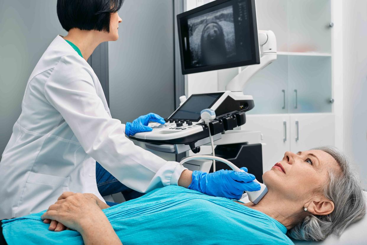
Ultrasound is a type of imaging technology that is used to diagnose, monitor, and treat a variety of medical conditions. It uses sound waves to generate live images (sonograms) of your organs, tissues, and fluids. It can also be used to change or destroy cells, like those that make up tumors.
Unlike imaging tests like positron emission testing (PET) scans and computed tomography (CT) scans, ultrasound does not involve radiation. It also does not typically require preparation or post-procedural instructions.
A healthcare provider, like an OB-GYN (a doctor who specializes in reproductive health) or cardiologist (a doctor who specializes in the heart), might perform an ultrasound scan. It can also be performed by an ultrasound technician known as an ultrasonographer. In that case, a radiologist (a doctor who specializes in medical imaging) will usually interpret your test results and send them to the provider who ordered your scan.
Key Terms
Sonography and ultrasound are often used interchangeably. However, there is a slight difference between these key terms. Here's what you need to know:
Sonography: The diagnostic medical test that uses ultrasound technology
Ultrasound: The technology that sonographers (people who specialize in sonography) use to generate medical images
Sonogram: The medical image that is produced after the completion of the ultrasound
Purpose
Your healthcare provider may recommend an ultrasound scan if you experience symptoms such as pain, swelling, or an unknown infection.
Here are some common ultrasounds and what they monitor:
Abdominal: Abdominal tissues and organs
Breast: Breast tissue
Echocardiogram: Heart
Obstetric: Fetus (infant developing in the uterus)
Ophthalmic: Ocular (eye) structures
Doppler: Blood flow through blood vessels and organs
Doppler fetal heart rate monitors: Fetal heartbeat
Bone sonometry: Bone fragility
Ultrasounds may also be used for:
Viewing blockages or growths in organs like the heart, liver, gallbladder, kidneys, bladder, or thyroid
Identifying conditions like ovarian cysts (fluid-filled sacs that develop inside or on the ovaries), uterine fibroids (non-cancerous growths in and around the uterus), and tumors
Guiding a needle into the body when taking a biopsy (sample) of cells or tissue
Ultrasound scans cannot adequately create images of all body parts. For example, it's difficult to examine areas of the body with a lot of air or gas, like the intestines and lungs. It's also difficult to see inside bones or view internal structures just under bones.
Ultrasound scans are routinely performed during pregnancy. They may be used early on for pregnancy dating. An anatomy ultrasound is typically performed between weeks 18-22 sometimes throughout a pregnancy to monitor fetal growth and development, including possible birth defects. They detect the fetus's heartbeat and provide images of the fetus.
Ultrasound can also determine placental location.. The placenta is the organ that delivers oxygen and nutrients to the fetus. A placenta positioned low in the uterus or over the cervix can potentially lead to serious complications for both the baby and the pregnant person.
Types of Ultrasound
Ultrasound falls into two main categories: diagnostic and therapeutic.
Diagnostic Ultrasound
Diagnostic ultrasound uses sound waves to create images of specific body parts. Probes may be placed on the skin or in the body.
Diagnostic ultrasound includes functional and anatomical ultrasounds:
An anatomical ultrasound creates images of internal structures. It might be used to examine organs like the brain and heart, muscles, or skin.
A functional ultrasound creates “information maps” by combining images with things like the movement of blood and tissue hardness or softness.
A Doppler ultrasound is a type of functional ultrasound. It can be used for several diagnostic reasons, including to measure blood flow through the heart as well as to monitor a fetus.
Therapeutic Ultrasound
Therapeutic ultrasound uses sound waves to change or destroy tissue, including tumors. It does not create images. Like other types of ultrasound, it's non-invasive and it doesn’t leave any wounds or scars.
How Does It Work?
Ultrasound works by producing high-frequency sound waves that humans can't detect. Sound waves are generated by a probe (transducer), which your healthcare provider places on your skin or inserts internally.
Internal ultrasounds include:
Transesophageal echocardiogram: A probe inserted into the esophagus to see the heart
Transrectal ultrasound: A probe inserted into the rectum, usually to see the prostate
Transvaginal ultrasound: A probe inserted into the vagina to see the uterus and ovaries
The sound waves transmit echoes back to the probe and generate an image in the video monitor.
Ultrasound images can be affected by:
Volume (amplitude) of the sound waves
Pitch (frequency) of the sound waves
How long it takes the sound waves to travel
Body structure or tissue the sound waves travel through
Your healthcare provider will usually apply an ultrasound gel to your skin before using the probe. The gel improves the image quality by helping the ultrasound waves pass into your body.
Some ultrasound devices are more transportable. For example, hand-held ultrasound machines can connect to a cell phone or tablet. Providers can use this “point of care ultrasound” (POCUS) in their offices or at a patient’s bedside, allowing them to quickly identify potentially serious conditions.
Before the Test
An ultrasound procedure does not usually require special preparations. You may be asked to wear loose-fitting clothing or change into a gown to expose the area of the body being examined.
You will likely fill out a questionnaire before the scan, including questions about your family and medical history as well as your current medications. The provider performing your scan will review the questionnaire with you before beginning.
You may be asked to refrain from eating or drinking before a scan. If you're having a bladder ultrasound, you may need to drink more water.
The duration of your ultrasound can vary depending on the area examined. Most exams take about 30 minutes, but it might take up to an hour. You will typically not require sedation or anesthesia as the procedure is not painful.
During the Test
You’ll likely lie face-up on an examination table for the ultrasound, but your positioning may depend on the area examined. Your healthcare provider will apply ultrasound gel to the transducer or to your body. The gel may feel cold, but it should not be painful. You can let the provider know if you feel too much pressure or any pain.
If you’re getting a diagnostic ultrasound, the provider will move the ultrasound probe over the area, adjusting for depth and contrast in order to create the clearest images possible. The transducer will instantly send images to an imaging screen and save the images for later review on a computer. You may or may not be able to see them during the procedure. The provider may also print images from the ultrasound machine.
After the Test
Once you complete the test, your healthcare provider will wipe off any remaining ultrasound gel and provide further instructions. They will give you privacy if you need to change back into your clothes.
You will probably be able to drive yourself home and resume all normal activities.
Risks and Precautions
Ultrasound is non-invasive and not usually painful. However, pressure from the transducer might create some discomfort, especially if the area examined is painful or sensitive.
Unlike other imaging studies, such as X-rays or a CT scan, ultrasound does not involve using radiation. It’s generally considered safe for children, older adults, and pregnant persons. However, it can affect body fluids and tissues. For example, ultrasound can slightly heat tissues or create small gas pockets. It should only be used when necessary for gaining medical information.
How to Prepare for an Ultrasound
An ultrasound scan does not usually require any preparation. As with all testing, you will need to consider the following:
Location: Because ultrasounds are very portable, the location where the ultrasound scan is performed can vary widely. For example, you may have it at a doctor's office, imaging center, hospital, or urgent care center.
Food and drink: Your healthcare provider will instruct you on whether you need to refrain from eating or drinking before the exam.
Medications: You can usually take your medications as prescribed before an ultrasound.
What to bring: Bring your identification card (such as a driver's license) and insurance card to the testing center. Your healthcare provider may give you a form called an order that describes the imaging tests being performed. They may also send an order in advance.
Emotional support: You might be able to bring someone with you, depending on the ultrasound type and location. Ask if this is allowed when you schedule the appointment.
Your insurance company will likely cover a portion of the costs if your ultrasound is medically necessary. Contact your insurance company before the procedure to see what they cover and if there are any payment plan options. The imaging location might also offer a discount if you pay in cash.
Related: What Should a Medical Scan Cost?
Results
Some healthcare providers performing an ultrasound procedure will interpret the images themselves. For example, an OB-GYN will take an ultrasound and review images of a fetus. A radiologist might examine the images if the ultrasound scan was performed by a technician. In this case, you may need to wait several days for the results.
You may receive your results via in-office appointment, mobile app, or over the phone, depending on the reasons for the ultrasound and the results.
Interpreting Your Results
You may need a follow-up ultrasound to better visualize certain areas or evaluate treatment responses. If you are pregnant, you might have multiple ultrasounds throughout your pregnancy, especially if your OB-GYN is monitoring a specific condition or possible concern.
Ultrasound results depend on the quality of the images, as well as the area examined. They may help your healthcare provider diagnose a condition or decide which further testing to recommend.
Ultrasound can be one part of a group of tests that include blood tests and additional imaging procedures like an X-ray or CT scan. Additional testing can potentially help your healthcare provider make the most accurate diagnosis or rule out other conditions.
A Quick Review
Ultrasound is a technology that uses sound waves to diagnose, monitor, and treat a variety of conditions, including pregnancy. Diagnostic ultrasound provides images of the area examined, and therapeutic ultrasound changes or destroys tissue, like tumors.
Ultrasound is a generally safe tool for gaining medical information because it doesn't use radiation. A scan is painless and usually takes about 30 minutes. You'll likely be able to resume normal activities immediately afterward.
Ultrasound has limitations. For example, it's difficult to view inside intestines, lungs, and bones. Therefore, healthcare providers may order additional imaging studies or testing.
For more Health news, make sure to sign up for our newsletter!
Read the original article on Health.
