An Overview of Retinal Tear

Medically reviewed by Johnstone M. Kim, MD
A retinal tear is a common age-related eye condition. It causes eye floaters and flashes of light and may lead to more severe vision problems.
On its own, a torn retina will not damage your eyesight. However, prompt treatment is needed. A retinal tear can quickly progress to a retinal detachment, which can cause permanent vision loss.
This article discusses retinal tears. It examines the symptoms, causes, and complications that can occur from a torn retina. It also explains when to see a vision specialist and how a torn retina is treated.

What Is a Retinal Tear?
The retina is a thin layer of light-sensitive tissue that lines the back of the eye. Located near the optic nerve, it receives light and sends pictures to the brain of what the eye sees.
As we age, the vitreous gel, a clear jelly-like substance that fills most of the eye’s interior, begins to break down, shrink, and pull away from the retina. This process is known as posterior vitreous detachment.
Most of the time, this happens without causing problems. Sometimes, though, the vitreous sticks to the retina and pulls it hard enough to cause a hole or tear.
Fluid can leak through the tear and cause the retina to lift away from the back of the eye, known as a retinal detachment. When this happens, the retina is unable to receive and process light. As a result, it sends distorted images to the brain—or none at all.
Retinal Tear vs. Retinal Hole
A retinal hole is a retinal tear that occurs in or near the center of the retina in an area known as the macula. Also called a macular hole, it may be more noticeable than a tear in the outer parts of the retina.
A retinal hole distorts your central vision, causing things to look blurry, cloudy, or wavy and making it difficult to see smaller details. Left untreated, the hole will grow, casting a dark shadow or blindspot in one eye.
Retinal Tear Symptoms
A torn retina is not painful, and you may not initially have any apparent signs. Symptoms of a retinal tear include flashes and floaters:
Flashes look like flashing lights, streaks of lightning, or stars in your field of vision. Flashes are an optical illusion caused by the vitreous pulling on the light-sensitive retina.
Floaters are small dark shapes that float across your vision. They can look like spots, threads, squiggly lines, or even little cobwebs. Eye floaters are often more noticeable when looking at a plain surface.
Complications
Retinal tears can quickly progress into the following more serious eye problems.
Vitreous Hemorrhage
A retinal tear can cause blood to leak into the vitreous, known as a vitreous hemorrhage.
A vitreous hemorrhage is not painful but does cause:
An increase in cobweb-like floaters
Hazy vision
Seeing shadows
A red hue or tint to vision in that eye
A vitreous hemorrhage may clear on its own but can also cause further damage to your eye and should be examined.
Retinal Detachment
Retinal tears can quickly progress to retinal detachment and cause the following symptoms:
Flashes of light in the eye (most common)
Visible spots called floaters (most common)
A sudden increase in size and number of floaters, indicating a retinal tear may be occurring
A sudden appearance of light flashes, which could be the first stage of a retinal tear or detachment
Having a shadow appear in your peripheral (side) field of vision
Seeing a gray curtain slowly moving across your field of vision
Experiencing a sudden decrease in sight, including focusing trouble and blurred vision
Having a headache
Seek Prompt Medical Care
A retinal tear can quickly cause a retinal detachment, which is a medical emergency. Call your eye doctor immediately if you experience the following:
A rapid increase in floaters or flashers
A shadow or curtain over your field of vision
Sudden trouble focusing or blurred vision
A delay in treatment could worsen your outcome.
What Causes a Retinal Tear?
The retina processes light through light-sensitive cells called photoreceptor cells. These cells detect light stimuli, which are interpreted as images.
The photoreceptor cells pass the information on to the optic nerve, which sends visual information to the brain. The brain then sorts through the information and “develops” the pictures.
In most cases, a retinal tear occurs when the vitreous gel inside the eye, also called the vitreous humor, contracts and tears the retina away from the eye wall.
The primary function of vitreous gel is to help the eyeball hold its spherical shape during fetal eye development. After the eye develops in utero, the purpose of the vitreous gel is unknown. There is still a lot to learn about the role of the gel.
This gel also helps the retina hold its place against the interior wall of the eyeball. The contraction of the vitreous gel can occur slowly over time or suddenly after experiencing trauma to the eye.
Risk Factors for Retinal Tears
While a retinal tear can occur in anyone at any age, they become more common after age 60. Other risk factors for retinal tears include:
Family history of retinal tears or detachment
Nearsightedness
Previous serious eye injury
Prior eye surgery, such as cataract or glaucoma surgery
A retinal tear or detachment in the other eye
Use of medications to constrict pupils, such as pilocarpine, which treats glaucoma age-related blurry near vision (presbyopia)
Weak areas of the retina (retinal degeneration)
Some health conditions can also increase your risk of a retinal tear, such as:
Sickle cell disease
Inflammatory disorders
Autoimmune diseases
Certain cancers
Certain hereditary eye conditions
Retinopathy of prematurity
Diagnosis
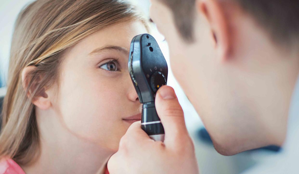
Retinal tears can be diagnosed during an eye exam by an optometrist or ophthalmologist.
Retinal tears are diagnosed through ophthalmoscopy. This painless test uses an ophthalmoscope to magnify and illuminate the eye. This allows for a closer inspection of the retina and other structures in the eye.
Ophthalmoscopes come in three different types, which your eye doctor may use:
Direct ophthalmoscopes are handheld, like a flashlight.
Indirect ophthalmoscopes are worn over the doctor's eyes like a miner's helmet.
Slit lamps are attached to a table by a brace where you rest your chin and forehead.
You may be given eye drops to dilate your pupils prior to the test. The drops take 20 to 30 minutes to work and may cause temporary light sensitivity.
Other tests may be needed, particularly if bleeding obstructs the view of the retina. Your eye doctor may also perform the following:
An ultrasound, which uses sound waves that bounce off the back of the eye to create a better picture of the retina
Optical coherence tomography (OCT), which uses light waves to take a cross-sectional image of the eye
Related: What to Expect From an Eye Exam
Treatment
A retinal tear does not always require treatment. Low-risk tears with no symptoms can sometimes be monitored closely without treatment. Some tears even resolve on their own.
In most cases, though, surgery is needed to reseal the retina. Treatment can often be done in an ophthalmologist's office and may include:
Laser surgery (photocoagulation): Your healthcare provider will use a laser to make small burns around the retinal tear. The scarring that results will seal the retina to the underlying tissue, helping to prevent a retinal detachment.
Freezing treatment (cryopexy): Your healthcare provider will use a special freezing probe to freeze the retina surrounding the retinal tear. The result is a scar that helps secure the retina to the eye wall.
Both procedures create a scar that seals the retina to the back of the eye, preventing fluid from traveling through the tear and under the retina. The procedure usually prevents the retina from detaching completely.
How Long Does It Take to Heal a Torn Retina?
Healing after treatment for a torn retina can take a while. After about 10 days, you should start to see improvement. However, it can take up to six months for the healing process to be complete.
Summary
A retinal tear can cause you to see stars, flashes of light, and specks or squiggles floating across your eye. The retina is a light-sensitive part of the eye that transmits images to the brain.
Most retinal tears are caused by the natural aging process and won’t damage your vision. However, it can lead to serious complications, including retinal detachment and vitreous hemorrhage.
A torn retina should be evaluated by an eye doctor. Small tears may heal on their own, but most retinal tears require surgery to prevent further damage and permanent vision loss.
With prompt treatment, retinal tears usually heal entirely. However, recovery can be slow, and it can take up to six months before your vision returns to normal.
